Research
- Research Areas
- Astrophysics & Cosmology (Observ.)
- Astrophysics & Cosmology (Theo.)
- Atomic Physics (Expt.)
- Atomic Physics (Theo.)
- Beam Physics
- Biological Physics
- Condensed Matter Physics (Expt.)
- Condensed Matter Physics (Theo.)
- General Relativity
- Microscopy
- Nuclear Physics
- Particle Physics (Expt.)
- Particle Physics (Theo.)
- Institutes & Centers
- Undergrad Research
- Graduate Research
Undergraduate Research Profiles
Below, you can read the profiles of a couple University of Chicago physics majors who have been active in research. A few past profiles are archived here. Enjoy…
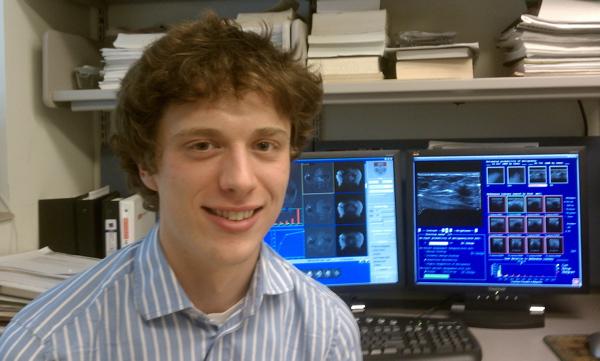 Jeremy Bancroft Brown
Undergraduate Student: Class of
2011
Major: Physics
Hometown: Amherst,
Massachusetts
Awards (HS): National Merit Commended
Student
Awards (Univ): Dean's List, Phi Beta Kappa, Class of
2011 Student Marshal, Grainger Senior Scholarship
(physics)
Research: Computer-aided Diagnosis, Atomic
Theory
Research Advisors: Maryellen
Giger, Lorenzo Curtis and David Ellis (Toledo)
Since October 2008, I have worked as a student researcher for Maryellen Giger, Ph.D. within the Department of Radiology and the interdisciplinary Graduate Programs of the Committee on Medical Physics. My research with Professor Giger is focused on computer-aided diagnosis (CAD) for a medical imaging modality known as dynamic contrast-enhanced magnetic resonance imaging (DCE-MRI). In recent years, DCE-MRI has become a widely used clinical tool for cancer diagnosis. In particular, it is used for surgical staging of newly diagnosed cancer, evaluation of cancer treatment, and screening of patients with known risk factors for developing cancer. At the University of Chicago, physicians most commonly employ DCE-MRI to diagnose and investigate carcinomas of the breast and prostate.
A DCE-MRI exam involves the acquisition of a three-dimensional image series over time (i.e., 4D data). During the exam, a pre-contrast 3D image series is first obtained. After this, the patient receives an intravenous dose of a paramagnetic contrast agent, and additional 3D images (with approximately millimeter spatial resolution) are subsequently acquired at multiple time points. The contrast agent reduces the longitudinal relaxation time (T1) of its surroundings, and can therefore be visualized by selecting a MRI pulse sequence that weights the image contrast towards the T1 values of the patient’s tissues. Additionally, because the contrast agent is initially localized to the blood pool, it primarily circulates to well-perfused areas of the body. This attribute is diagnostically useful, because tumors tend to be highly vascularized. Moreover, invasive cancers typically exhibit a disorganized, “leaky” vasculature (due to angiogenesis) that produces a characteristic rapid uptake and rapid washout of the contrast agent. In comparison, benign cell proliferation processes generally present with slower contrast uptake and washout.
In current clinical practice, radiologists are able to extract useful information from DCE-MRI examinations, and they routinely incorporate this information into cancer diagnosis and treatment decisions by collaborating with surgeons, oncologists, and pathologists. However, there is substantial room to improve the interpretation of DCE-MRI data. A typical DCE-MRI dataset comprises 6 to 50 timepoints each containing 60 to 200 image slices, which are each composed of at least 215 voxels. This large quantity of digital data is ideally suited to computerized analysis methods. The primary goals of CAD research for DCE-MRI in Professor Giger’s lab include accurate tumor detection, delineation, and characterization to aid doctors in biopsy and treatment decisions, as well as to extract quantitative information related to patient phenotypes, prognoses, and responses to therapy. The methods used combine machine learning (neural networks and artificial intelligence) techniques with knowledge of the NMR physics and pathophysiology that underlie the data.
My own research focus within Professor Giger’s lab has been on understanding kinetic patterns of contrast uptake and washout in DCE-MRI exams. One project that I have contributed to uses a pattern recognition algorithm known as fuzzy C-means (FCM) in order to identify characteristic kinetic behavior from a given tumor. Another strategy that I have investigated involves summarizing the four-dimensional (spatial and temporally indexed) DCE-MRI data using three-dimensional (spatially indexed) kinetic feature images, which can then be analyzed using fractal dimension formalisms and higher-order texture statistics. A third thrust of my research entails comparing these population data-driven image analysis techniques to methods that extract quantitative physiologic parameters from the DCE-MRI data using pharmacokinetic models. This research requires some use of computer simulations, but it relies heavily on retrospective clinical data from hundreds of patients. My research in Professor Giger’s lab has been supported by an American Association of Physicists in Medicine (AAPM) Summer Fellowship, a University of Chicago Biological Sciences Collegiate Division (BSCD) Summer Fellowship, and Professor Giger’s research grants from the National Cancer Institute (NCI) and the Department of Energy (DOE).
During the summer of 2008, I participated in the National Science Foundation (NSF) Research Experience for Undergraduates (REU) program at the University of Toledo under the guidance of Larry Curtis, Ph.D. and David Ellis, Ph.D. In my project there, we used a semi-empirical method to characterize the 3s23p2–3s3p3 J=2 transition array in singly-ionized phosphorous (P II). In this method, Slater, spin-orbit, and radial parameters were fitted to experimental energy levels in order to obtain a description of the array in terms of LS-coupling basis vectors. The various intermediate coupling (IC) and configuration interaction (CI) amplitudes resulting from this model were then used to predict the branching fractions of transitions within the array. It was then possible to compare the semi-empirical predictions to branching ratios measured using beam-foil spectroscopy at the Toledo Heavy Ion Accelerator (THIA) laboratory. This research was motivated by the fact that P II is common in interstellar clouds. Thus, precise laboratory measurement of its energy levels and oscillator strengths (together with theoretical modeling of those quantities in order to guide and inform the experimental work) facilitates quantification of its abundance. This knowledge in turn allows astrophysicists to better understand the chemical evolution of galaxies.